- Home
- The Clinic
- Pathologies
- Techniques / Technology
- INUSpheresis®
- Clinical Analysis
- Blog
- Contact
A type of immune cell that appears to block the progression of melanoma and other cancers in animal models has been identified by researchers.
Researchers at Massachusetts General Hospital (MGH) have identified a type of immune cell that appears to block the progression of melanoma and other cancers in animal models. These subcapsular sinus macrophages (SCS) form a protective layer around lymph nodes, preventing the entry of tiny structures that carry bits of tumor tissue and help cancer grow and spread. However, the SCS macrophage barrier appears to be temporary, as it breaks down as the tumor progresses and in response to some cancer treatment drugs.
Macrophages found in tumors are typically viewed as promoters of cancer growth, for example, by helping to form new blood vessels that deliver nutrients to tumor cells, says Mikael Pittet, PhD, of the MGH Center for Systems Biology, who led the study published April 8, 2016, in the journal Science. My lab studies how tumors communicate with the immune system throughout the body, and we are particularly interested in whether tumors also interact with macrophages that reside outside the tumor.
One potential means by which molecular signals could be transferred from tumors toimmune cells are small membrane compartments called tumor-derived extracellular vesicles (tEVs), which are known to bind to and activate many different types of cells. Measuring tEV levels can be used to predict treatment response and survival, but assessing the impact of tEVs in living animals has been difficult. The MGH team combined genetic and scanning approaches in a novel way to track tEVs and their targets.
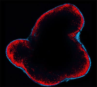
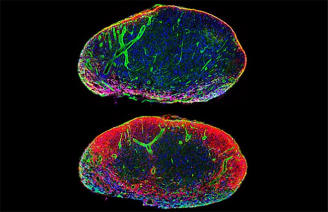
In mice with tumor cells genetically modified to produce tEVs with a light-emitting marker, the researchers confirmed that tEVs can leave tumors and travel through the body, finding that they were most highly concentrated in lymph nodes, to which they were transported via lymphatic vessels. In another group of mice with melanomas carrying different informant proteins, the team found that tEVs interact primarily with SCS macrophages, which form a layer directly on the fibrous capsule surrounding the lymph nodes.
To determine whether this observation in mice was relevant to human disease, the researchers examined cancer-free sentinel lymph nodes —the ones closest to the tumor to which the tumor would be expected to spread first—in 13 patients with melanoma. Although the nodes themselves were confirmed to be free of melanoma, melanoma-derived material was found in the SCS macrophages surrounding the nodes in 90 percent of patients. The presence of tumor-derived material in these macrophages did not reflect how far the primary tumor had progressed.
Other experiments found that SCS macrophages act as tumor suppressors in two mouse melanoma models and in a lung cancer model. This is in stark contrast to macrophages within tumors, which normally promote cancer. While the current study demonstrated that SCS macrophages suppress cancer by limiting the spread of tEVs, the density of SCS macrophages around lymph nodes begins to decrease as tumors grow. Treatment with chemotherapy and immune therapy drugs was also found to disrupt the SCS macrophage barrier. Once tEVs enter the lymph nodes, they bind to B cells, which produce antibodies that accelerate tumor growth.
Pittet, an associate professor of radiology at Harvard Medical School, states: "Given the interest in developing therapies that extinguish tumor-promoting macrophages within tumors, it may be useful to determine whether these treatments also affect protective SCS macrophages. The best outcome would likely be to eliminate the activities of tumor-promoting macrophages within tumors while preserving the activities of tumor-suppressing SCS macrophages. It would also be useful to determine whether SCS macrophages can be reinforced to prevent the delivery of tEVs to lymph nodes and to better understand how tEV-activated B cells promote cancer growth.
Source: Science Daily